Biology · Cell: Structure and Function · Chapter notes
Cell: The Unit of Life · Class 11 NEET Notes
Cell: The Unit of Life Class 11 notes for NEET: 11 drawn diagrams, prokaryotic vs eukaryotic cells, every organelle, 14 worked questions and a fact sheet.
Every Class 11 and 12 chapter in the app: a game to play, a short read, then questions that check you. Free to start

In short
Cell: The Unit of Life is NCERT Class 11 Biology Chapter 8. It covers cell theory, prokaryotic and eukaryotic cells, and every organelle. In NEET it gives about 4 to 5 questions a year. Prokaryotic cell structure is the most tested topic, and mesosome functions are the single most repeated concept.
Contents
- ·How to Read This Set and what NEET 2026 asks
- 01What Is a Cell, and Cell Theory who added what
- 02An Overview of Cells: Size and Shape three superlatives, three cells
- 03Prokaryotic Cells the most tested topic
- 04Eukaryotic Cells: Plant vs Animal four differences
- 05The Cell Membrane fluid mosaic and transport
- 06The Cell Wall primary, secondary, middle lamella
- 07The Endomembrane System what is in, what is out
- 08Mitochondria and Plastids the two semi-autonomous organelles
- 09Ribosomes and the Cytoskeleton why S values do not add
- 10Cilia, Flagella, Centrosome and Centrioles doublets vs triplets
- 11The Nucleus and Chromosomes pores, nucleolus, centromere
- 12Microbodies two lines, one possible question
- 13Quick Review: What NEET Repeats 22 cards for the last week
- ★Cell: The Unit of Life · Fact Sheet
How to Read This Set
- Read each section once, slowly. Then look at its diagram and name every part without the labels.
- Orange boxes are traps. NEET loves them. Read every one.
- Questions after each section are NEET level. Try each one before reading the answer.
- The Quick Review at the end lists what NEET repeats. Use it in the last week, not the first.
- The Fact Sheet is one page. Keep it for the night before the exam.
The NMC syllabus for NEET UG 2026 starts with cell theory and the cell as the basic unit of life. It then lists prokaryotic and eukaryotic cells, and plant and animal cells. It names the cell envelope, cell membrane and cell wall. It covers the structure and function of organelles.
Organelles named are ER, Golgi bodies, lysosomes, vacuoles, mitochondria, ribosomes, plastids and microbodies. Cytoskeleton, cilia, flagella, centrioles and the nucleus are also included. These notes cover all of it, and nothing outside NCERT.
We analysed 55 NEET questions from 2015 to 2025. This chapter gave about 4 to 5 questions a year. Prokaryotic cell structure was the most tested topic.
What Is a Cell, and Cell Theory
Points
- A cell is the smallest unit that can live on its own.
- A one-celled organism does every job of life inside that single cell.
- Anything less than a full cell cannot live independently.
- Antonie Von Leeuwenhoek first saw and described a live cell.
- Robert Brown later discovered the nucleus.
Who added what
- Matthias Schleiden (1838), a German botanist. He studied many plants. He saw that all plants are made of different kinds of cells. These cells form the tissues of the plant.
- Theodore Schwann (1839). He studied many animal cells. He reported a thin outer layer on cells, now called the plasma membrane.
- Schwann also concluded that the cell wall is unique to plant cells.
- He proposed that the bodies of animals and plants are made of cells and products of cells.
- Schleiden and Schwann together formulated the cell theory.
- Their gap: the theory did not explain how new cells are formed.
- Rudolf Virchow (1855) explained it. Cells divide, and new cells form from pre-existing cells.
- He wrote it as Omnis cellula-e cellula.
- Virchow modified the cell theory into its final form.
Cell theory as understood today
- All living organisms are composed of cells and products of cells.
- All cells arise from pre-existing cells.
- "Cells arise from pre-existing cells" is Virchow's idea, not Schleiden's or Schwann's. NEET has tested this exact credit swap more than once.
- The current NCERT calls Schwann "a German Zoologist". Older editions said British. In the exam, go with the current NCERT sentence. It is the source NEET follows.
Assertion (A): The cell theory of Schleiden and Schwann could not explain how new cells are formed.
Reason (R): Rudolf Virchow showed that cells divide and new cells form from pre-existing cells.
- Both A and R are true and R is the correct explanation of A
- Both A and R are true but R is not the correct explanation of A
- A is true but R is false
- A is false but R is true
Show answer
An Overview of Cells: Size and Shape
Points
- Onion cells, a plant cell, have a distinct cell wall outside the cell membrane.
- Human cheek cells, an animal cell, have only an outer membrane as the boundary.
- Inside each cell is a dense, membrane-bound structure called the nucleus.
- The nucleus holds the chromosomes, which carry the genetic material, DNA.
- Eukaryotic cells have a membrane-bound nucleus.
- Prokaryotic cells lack a membrane-bound nucleus.
- In both, a semi-fluid matrix called cytoplasm fills the cell.
- Cytoplasm is the main arena of cell activity in plant and animal cells.
- Eukaryotic cells also have membrane-bound organelles: ER, Golgi complex, lysosomes, mitochondria, microbodies and vacuoles.
- Prokaryotic cells lack such membrane-bound organelles.
- Ribosomes are non-membrane-bound. They occur in all cells, prokaryotic and eukaryotic.
- In eukaryotes, ribosomes are found in the cytoplasm, chloroplasts, mitochondria and on rough ER.
- Animal cells have another non-membrane-bound organelle, the centrosome, which helps in cell division.
Size
| Cell | Size given in NCERT |
|---|---|
| Mycoplasma (smallest cells) | 0.3 µm in length |
| Bacteria | 3 to 5 µm |
| Human red blood cell | about 7.0 µm in diameter |
| Ostrich egg | largest isolated single cell |
| Nerve cells | some of the longest cells |
Shape
- Cells may be disc-like, polygonal, columnar, cuboid, thread-like or irregular.
- Shape varies with function.
- "Smallest cell" is Mycoplasma at 0.3 µm. "Largest single cell" is the ostrich egg. "Longest cells" are nerve cells. Three superlatives, three different cells.
A student places Mycoplasma cells in a straight line, end to end, across one human red blood cell. Use NCERT sizes. About how many Mycoplasma cells fit across the RBC's diameter?
- 0.04
- 2
- 23
- 230
Show answer
Prokaryotic Cells
Who they are
- Prokaryotic cells are bacteria, blue-green algae, mycoplasma and PPLO (Pleuro Pneumonia Like Organisms).
- They are usually smaller than eukaryotic cells.
- They multiply more rapidly.
- They vary greatly in shape and size.
- Bacteria have four basic shapes: bacillus (rod), coccus (spherical), vibrio (comma) and spirillum (spiral).
Inside the cell
- All prokaryotes have a cell wall around the cell membrane. Mycoplasma is the exception.
- The fluid matrix filling the cell is the cytoplasm.
- There is no well-defined nucleus.
- The genetic material is basically naked. It is not enveloped by a nuclear membrane.
- Many bacteria also have small circular DNA outside the genomic DNA. These are plasmids.
- Plasmids give bacteria special traits. One example is resistance to antibiotics.
- Plasmid DNA is used to monitor bacterial transformation with foreign DNA. (You will meet this again in Biotechnology.)
- There are no membrane-bound organelles like those in eukaryotes. Ribosomes are present.
The mesosome
- A mesosome is a special membranous structure. It forms when the cell membrane folds inward.
- It takes the form of vesicles, tubules and lamellae.
- Functions: cell wall formation, DNA replication and distribution to daughter cells, respiration and secretion.
- It also increases the surface area of the plasma membrane and its enzymatic content.
- A mesosome is an infolding of the plasma membrane. It is not a separate membrane-bound organelle. Mesosome functions are the single most repeated concept in this chapter: 5 questions in our data.
- In some prokaryotes like cyanobacteria, membranous extensions into the cytoplasm contain pigments. These are chromatophores.
The cell envelope
- Most prokaryotes, especially bacteria, have a chemically complex cell envelope.
- It has three tightly bound layers, from outside in:
- Glycocalyx, the outermost layer
- Cell wall
- Plasma membrane
- Each layer has a distinct function, but together they act as one protective unit.
- The glycocalyx differs between bacteria in composition and thickness.
- A loose sheath is called a slime layer.
- A thick, tough layer is called a capsule.
- The cell wall decides the cell's shape. It gives strong structural support so the bacterium does not burst or collapse.
- The plasma membrane is semi-permeable. It interacts with the outside world.
- It is structurally similar to the membrane of eukaryotes.
- Gram staining. Bacteria differ in their cell envelopes and in how they respond to Gram's stain.
- Bacteria that take up the stain are Gram positive.
- Those that do not are Gram negative.
This chapter is a set of short games in the Logic Bloom app.
Surface structures
- Motile bacteria have thin filamentous extensions called flagella.
- A bacterial flagellum has three parts: filament, hook and basal body.
- The filament is the longest part. It extends from the cell surface outward.
- Pili are elongated tubular structures made of a special protein.
- Fimbriae are small bristle-like fibres sprouting out of the cell.
- In some bacteria, fimbriae help attach to rocks in streams and to host tissues.
- Pili and fimbriae do not help in movement. Only flagella do.
Ribosomes and inclusion bodies
- Prokaryotic ribosomes are associated with the plasma membrane.
- Each is about 15 nm by 20 nm in size.
- Each has two subunits, 50S and 30S, which together form a 70S ribosome.
- Ribosomes are the site of protein synthesis.
- Several ribosomes may attach to a single mRNA and form a chain called a polyribosome or polysome.
- The ribosomes of a polysome translate the mRNA into proteins.
- Reserve material is stored in inclusion bodies.
- Inclusion bodies are not bound by any membrane. They lie free in the cytoplasm.
- Examples: phosphate granules, cyanophycean granules and glycogen granules.
- Gas vacuoles are found in blue-green, purple and green photosynthetic bacteria.
Read these statements about mesosomes.
- (a) They are infoldings of the plasma membrane.
- (b) They help in cell wall formation.
- (c) They help in DNA replication and its distribution to daughter cells.
- (d) They are bounded by a double membrane, like mitochondria.
- (e) They increase the surface area of the plasma membrane.
Choose the correct answer from the options given below.
- (a), (b), (c) and (e) only
- (a), (b), (c) and (d) only
- (b), (c), (d) and (e) only
- (a) and (d) only
Show answer
| Column I | Column II |
|---|---|
| A. Glycocalyx as a loose sheath | I. Attachment to rocks in streams and host tissues |
| B. Cell wall | II. Slime layer |
| C. Fimbriae | III. Stores reserve material without a membrane |
| D. Inclusion body | IV. Decides cell shape and prevents bursting |
Choose the correct answer from the options given below.
- A-II, B-IV, C-III, D-I
- A-IV, B-II, C-I, D-III
- A-II, B-IV, C-I, D-III
- A-I, B-IV, C-II, D-III
Show answer
Eukaryotic Cells: Plant vs Animal
Points
- Eukaryotic cells are found in protists, plants, animals and fungi.
- Membrane-bound organelles divide the cytoplasm into compartments.
- They have an organised nucleus with a nuclear envelope.
- They have a complex locomotory and cytoskeletal structure.
- Their genetic material is organised into chromosomes.
Plant cell vs animal cell
| Feature | Plant cell | Animal cell |
|---|---|---|
| Cell wall | Present | Absent |
| Plastids | Present | Absent |
| Large central vacuole | Present | Absent |
| Centrioles | Absent in almost all plant cells | Present |
- NCERT says centrioles are absent in almost all plant cells, not all. An option saying "never present in any plant cell" is an absolute claim. Treat such options with suspicion.
The Cell Membrane
Composition
- Its detailed structure was studied after the electron microscope arrived in the 1950s.
- Chemical studies used human red blood cells (RBCs).
- They showed the membrane is mainly lipids arranged in a bilayer.
- The polar heads face the outer sides. The hydrophobic tails face the inside.
- So the non-polar tails stay protected from the watery surroundings.
- The lipids are mainly phosphoglycerides.
- The membrane also has cholesterol.
- It also has proteins and carbohydrates.
- The ratio of protein to lipid varies in different cell types.
- In the human erythrocyte membrane: about 52% protein and 40% lipids.
- Peripheral proteins lie on the membrane surface.
- Integral proteins are partly or totally buried in the membrane.
The fluid mosaic model
- Proposed by Singer and Nicolson (1972). It is the widely accepted model.
- The lipid is quasi-fluid in nature.
- This lets proteins move sideways within the overall bilayer.
- This ability to move is called the membrane's fluidity.
- Fluidity matters for cell growth, intercellular junctions, secretion, endocytosis and cell division.
A membrane is a fixed, rigid wall.
Its lipids are quasi-fluid. Proteins drift sideways through the bilayer, which is exactly why it is called "fluid".
Transport across the membrane
- The membrane is selectively permeable. Some molecules cross it; others do not.
- Passive transport: molecules move without using energy.
- Neutral solutes may cross by simple diffusion, from higher to lower concentration.
- Water also moves by diffusion, from higher to lower concentration. Movement of water by diffusion is osmosis.
- Polar molecules cannot pass through the non-polar lipid bilayer. They need a carrier protein.
- Active transport: a few ions or molecules move against the concentration gradient.
- This uses energy from ATP. Example: the Na⁺/K⁺ pump.
Statement I: Polar molecules cross the lipid bilayer easily by simple diffusion.
Statement II: The Na⁺/K⁺ pump moves ions against their concentration gradient using ATP.
- Both Statement I and Statement II are true
- Both Statement I and Statement II are false
- Statement I is true but Statement II is false
- Statement I is false but Statement II is true
Show answer
Read less, play more
Now play it.
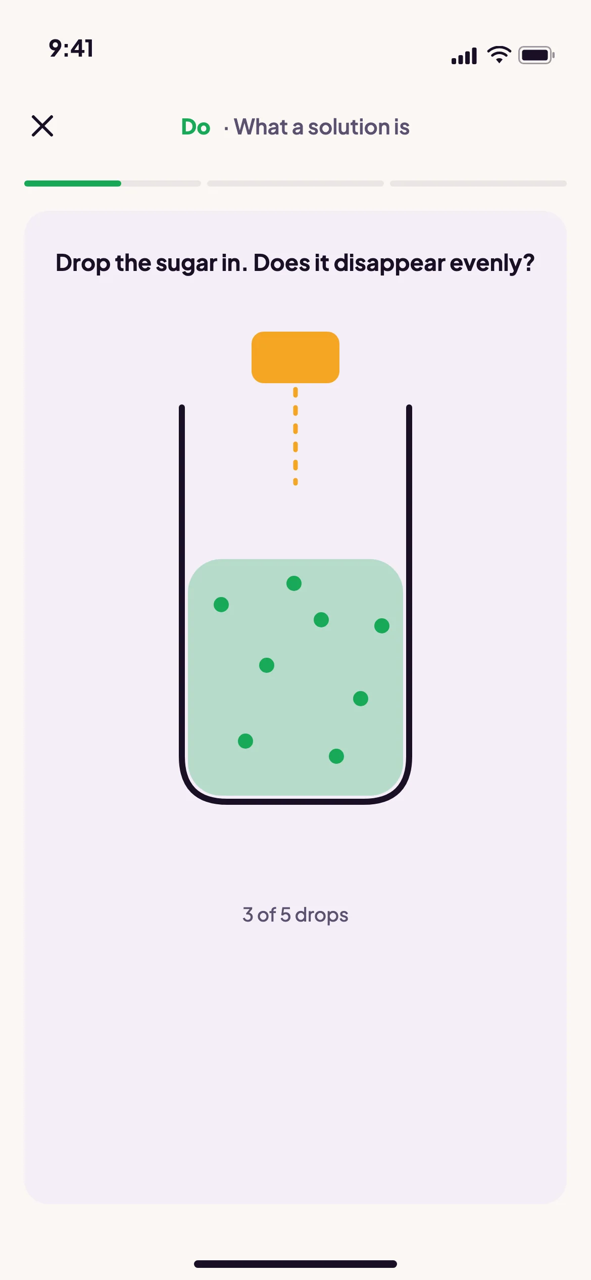
In the app a chapter starts as a game you play, then a short read, then questions that check you. A full chapter, understood, in 45 to 60 minutes.
The Cell Wall
Points
- The cell wall is a non-living, rigid structure.
- It covers the plasma membrane of fungi and plants from outside.
- It gives shape to the cell and protects it from mechanical damage and infection.
- It helps in cell-to-cell interaction.
- It is a barrier to unwanted macromolecules.
- Algal cell wall: cellulose, galactans, mannans and minerals like calcium carbonate.
- Other plants: cellulose, hemicellulose, pectins and proteins.
- The primary wall belongs to a young plant cell and can grow.
- This growth slowly stops as the cell matures.
- The secondary wall then forms on the inner side, towards the membrane.
- The middle lamella is a layer mainly of calcium pectate. It holds neighbouring cells together.
- Plasmodesmata cross the cell wall and middle lamella. They connect the cytoplasm of neighbouring cells.
- The secondary wall forms on the inner side, between the primary wall and the membrane. Students often place it outside, because "secondary" sounds like "added on top".
Which of these statements about the plant cell wall are correct?
- (a) The middle lamella is made mainly of calcium pectate.
- (b) The secondary wall forms on the outer side of the primary wall.
- (c) The primary wall of a young cell is capable of growth.
- (d) Plasmodesmata connect the cytoplasm of neighbouring cells.
- (e) Algal cell walls may contain calcium carbonate.
Choose the correct answer from the options given below.
- (a), (b) and (d) only
- (b), (c) and (e) only
- (a), (c) and (d) only
- (a), (c), (d) and (e) only
Show answer
The Endomembrane System
What is in it, and what is not
- Many membranous organelles are distinct in structure and function.
- Some of them have coordinated functions. These form the endomembrane system.
- Included: endoplasmic reticulum (ER), Golgi complex, lysosomes and vacuoles.
- Not included: mitochondria, chloroplasts and peroxisomes.
- Why? Their functions are not coordinated with the four above.
- "Membrane-bound" does not mean "endomembrane". Mitochondria and chloroplasts have two membranes, yet they are outside the system. Peroxisome exclusion has appeared as a NEET question on its own.
This chapter is a set of short games in the Logic Bloom app.
Endoplasmic reticulum (ER)
- The ER is a network of tiny tubules scattered in the cytoplasm.
- It divides the space inside the cell into two compartments.
- Luminal: inside the ER.
- Extra luminal: the cytoplasm.
- ER with ribosomes on its outer surface is rough ER (RER).
- ER without ribosomes looks smooth. It is smooth ER (SER).
- RER is common in cells busy with protein synthesis and secretion.
- RER is extensive and continuous with the outer membrane of the nucleus.
- SER is the major site for lipid synthesis.
- In animal cells, lipid-like steroidal hormones are made in SER.
- Ribosomes make proteins, so RER is the protein line. SER has no ribosomes, so it makes lipids. Structure tells you function.
Golgi apparatus
- Camillo Golgi (1898) first saw densely stained net-like structures near the nucleus. These were later named Golgi bodies.
- They are many flat, disc-shaped sacs called cisternae.
- Each cisterna is 0.5 µm to 1.0 µm in diameter.
- The cisternae are stacked parallel to each other.
- The number of cisternae in a Golgi complex varies.
- They are arranged concentrically near the nucleus.
- The stack has a convex cis face (forming face) and a concave trans face (maturing face).
- The cis and trans faces are entirely different, but they are interconnected.
- Main job: packaging materials, for delivery inside the cell or for secretion outside it.
- Vesicles from the ER fuse with the cis face.
- The materials then move towards the trans face.
- So the Golgi stays closely associated with the ER.
- Proteins made by ribosomes on the ER are modified in the cisternae, then released from the trans face.
- The Golgi is the important site where glycoproteins and glycolipids are formed.
- ER vesicles enter at the cis face and leave from the trans face. Remember: cis comes in. In our data, Golgi glycoprotein and glycolipid formation was tested 4 times.
Lysosomes
- Lysosomes are membrane-bound vesicles formed by packaging in the Golgi.
- Isolated lysosomes are rich in almost all types of hydrolytic enzymes, the hydrolases: lipases, proteases and carbohydrases.
- These enzymes work best at acidic pH.
- They can digest carbohydrates, proteins, lipids and nucleic acids.
Vacuoles
- A vacuole is a membrane-bound space in the cytoplasm.
- It contains water, sap, excretory products and materials the cell does not need.
- It is bound by a single membrane called the tonoplast.
- In plant cells, vacuoles can occupy up to 90% of the cell's volume.
- In plants, the tonoplast moves ions and other materials against their concentration gradient into the vacuole.
- So their concentration is much higher in the vacuole than in the cytoplasm.
- In Amoeba, the contractile vacuole helps in osmoregulation and excretion.
- In many protists, food vacuoles form when they engulf food particles.
How many of the following are part of the endomembrane system?
Endoplasmic reticulum, Golgi complex, lysosome, vacuole, mitochondrion, chloroplast, peroxisome, ribosome
- Three
- Four
- Five
- Six
Show answer
Statement I: Vesicles from the ER fuse with the trans face of the Golgi apparatus.
Statement II: The Golgi apparatus is an important site for forming glycoproteins and glycolipids.
- Both Statement I and Statement II are true
- Both Statement I and Statement II are false
- Statement I is true but Statement II is false
- Statement I is false but Statement II is true
Show answer
In a plant cell, some ions are far more concentrated in the vacuole than in the cytoplasm. Which explains this best?
- Ions diffuse freely into the vacuole through the tonoplast.
- Water leaves the vacuole by osmosis, which concentrates the ions.
- The tonoplast transports ions against their concentration gradient into the vacuole.
- The contractile vacuole pumps ions into the central vacuole.
Show answer
Mitochondria and Plastids
Mitochondria
- Mitochondria are hard to see under the microscope unless specially stained.
- Their number per cell depends on the cell's physiological activity.
- They are sausage-shaped or cylindrical.
- Diameter: 0.2 to 1.0 µm (average 0.5 µm). Length: 1.0 to 4.1 µm.
- Each mitochondrion is a double membrane-bound structure.
- The outer and inner membranes divide its space into two watery compartments.
- The outer compartment.
- The inner compartment, called the matrix.
- The outer membrane forms a continuous boundary around the organelle.
- The inner membrane folds into the matrix. These folds are cristae.
- Cristae increase the surface area.
- Each membrane has its own specific enzymes.
- Mitochondria are the sites of aerobic respiration.
- They produce cellular energy as ATP. So they are called the "power houses" of the cell.
- The matrix has a single circular DNA molecule, a few RNA molecules and 70S ribosomes.
- It also has the components needed to make proteins.
- Mitochondria divide by fission.
Plastids
- Plastids are found in all plant cells and in euglenoides.
- They are easily seen under the microscope, as they are large.
- Each type bears specific pigments, which give plants their colours.
- There are three types by pigment:
| Type | What it holds | Example or role |
|---|---|---|
| Chloroplasts | Chlorophyll and carotenoid pigments | Trap light energy for photosynthesis |
| Chromoplasts | Fat-soluble carotenoids: carotene, xanthophylls | Yellow, orange or red colour |
| Leucoplasts | Colourless; store nutrients | See below |
- Leucoplasts by what they store:
- Amyloplasts store carbohydrates (starch). Example: potato.
- Elaioplasts store oils and fats.
- Aleuroplasts store proteins.
- Link each store to its word: amylo = starch, elaio = oil, aleuro = protein.
Chloroplasts in detail
- Most chloroplasts of green plants lie in the mesophyll cells of leaves.
- They may be lens-shaped, oval, spherical, discoid or ribbon-like.
- Length: 5 to 10 µm. Width: 2 to 4 µm.
- Their number varies. Chlamydomonas, a green alga, has one per cell.
- Mesophyll cells have 20 to 40 per cell.
- Chloroplasts are double membrane-bound.
- The inner membrane is relatively less permeable.
- The space inside the inner membrane is the stroma.
- The stroma holds flattened membranous sacs called thylakoids.
- Thylakoids are stacked like piles of coins. Each stack is a granum (plural: grana).
- Flat membranous tubules called stroma lamellae connect thylakoids of different grana.
- The thylakoid membrane encloses a space called the lumen.
- The stroma holds enzymes needed to make carbohydrates and proteins.
- It also holds small, double-stranded circular DNA and ribosomes.
- Chlorophyll pigments are in the thylakoids.
- Chloroplast ribosomes are 70S. They are smaller than cytoplasmic ribosomes, which are 80S.
Which features are common to both mitochondria and chloroplasts?
- (a) A double membrane
- (b) Their own circular DNA
- (c) 70S ribosomes
- (d) An inner membrane folded into cristae
- (e) Membership of the endomembrane system
Choose the correct answer from the options given below.
- (a), (b) and (c) only
- (a), (b), (c) and (d) only
- (a) and (d) only
- (b), (c) and (e) only
Show answer
Ribosomes and the Cytoskeleton
Ribosomes
- Ribosomes are granular structures.
- George Palade (1953) first saw them under the electron microscope as dense particles.
- They are made of RNA and proteins.
- They are not surrounded by any membrane.
- Eukaryotic ribosomes are 80S. Prokaryotic ribosomes are 70S.
- Each ribosome has two subunits, a larger one and a smaller one.
| Ribosome | Larger subunit | Smaller subunit |
|---|---|---|
| 80S (eukaryotic cytoplasm) | 60S | 40S |
| 70S (prokaryotes, mitochondria, chloroplasts) | 50S | 30S |
- "S" stands for the Svedberg unit. It is the sedimentation coefficient.
- It is an indirect measure of density and size.
50S + 30S = 80S, so S values must add up.
S measures how fast a particle settles, which depends on size and shape. So subunit values do not add. 50S and 30S make a 70S ribosome.
Cytoskeleton
- The cytoskeleton is a detailed network of filament-like protein structures in the cytoplasm.
- It has three parts: microtubules, microfilaments and intermediate filaments.
- Functions: mechanical support, motility, and keeping the cell's shape.
Read these statements about ribosomes.
- (a) A 70S ribosome is made of 50S and 30S subunits.
- (b) The S values of the two subunits add up to the S value of the whole ribosome.
- (c) Ribosomes in mitochondria and chloroplasts are 70S.
- (d) Several ribosomes on one mRNA form a polysome.
- (e) Ribosomes are bounded by a single membrane.
Choose the correct answer from the options given below.
- (a), (b) and (c) only
- (c), (d) and (e) only
- (a), (b), (c) and (d) only
- (a), (c) and (d) only
Show answer
Cilia, Flagella, Centrosome and Centrioles
Cilia and flagella
- Cilia and flagella are hair-like outgrowths of the cell membrane.
- Cilia are small. They work like oars and move the cell or the fluid around it.
- Flagella are longer. They move the cell.
- Prokaryotic flagella are structurally different from eukaryotic flagella.
- Electron microscopy shows each is covered by the plasma membrane.
- Its core is the axoneme. It has many microtubules running parallel to the long axis.
- The axoneme has nine pairs of doublets of peripheral microtubules, arranged in a ring.
- It also has a pair of centrally located microtubules.
- This is the 9+2 array.
- The central tubules are joined by bridges and enclosed by a central sheath.
- The central sheath connects to one tubule of each peripheral doublet by a radial spoke. So there are nine radial spokes.
- The peripheral doublets are also joined to each other by linkers.
- Both cilia and flagella emerge from centriole-like structures called basal bodies.
Centrosome and centrioles
- The centrosome usually contains two cylindrical structures called centrioles.
- Amorphous pericentriolar material surrounds them.
- The two centrioles lie perpendicular to each other.
- Each centriole looks like a cartwheel.
- It has nine evenly spaced peripheral fibrils of tubulin protein.
- Each peripheral fibril is a triplet. Adjacent triplets are linked.
- The central part of the proximal region is also protein. It is called the hub.
- The hub connects to the peripheral triplets through radial spokes made of protein.
- Jobs: centrioles form the basal body of cilia or flagella.
- They also form the spindle fibres of the spindle apparatus during cell division in animal cells.
This chapter is a set of short games in the Logic Bloom app.
- Axoneme ring: doublets. Centriole ring: triplets. The axoneme has two central microtubules; the centriole has a protein hub and no central microtubules.
| Column I | Column II |
|---|---|
| A. Peripheral ring of the axoneme | I. Proteinaceous hub |
| B. Peripheral ring of a centriole | II. Two microtubules in a central sheath |
| C. Centre of the axoneme | III. Nine doublets |
| D. Centre of a centriole (proximal region) | IV. Nine triplets |
Choose the correct answer from the options given below.
- A-III, B-IV, C-II, D-I
- A-IV, B-III, C-II, D-I
- A-III, B-IV, C-I, D-II
- A-IV, B-III, C-I, D-II
Show answer
The Nucleus and Chromosomes
Nucleus
- Robert Brown first described the nucleus, as early as 1831.
- Its material stains with basic dyes. Flemming named it chromatin.
- The interphase nucleus (a nucleus not dividing) has three things.
- Highly extended nucleoprotein fibres called chromatin.
- The nuclear matrix.
- One or more spherical bodies called nucleoli (singular: nucleolus).
- The nuclear envelope has two parallel membranes.
- The space between them is the perinuclear space, 10 to 50 nm wide.
- The envelope is a barrier between the nucleus and the cytoplasm.
- The outer membrane usually stays continuous with the ER. It also bears ribosomes.
- At many places, the two membranes fuse to form nuclear pores.
- RNA and protein molecules move both ways through these pores, between nucleus and cytoplasm.
- Normally, there is only one nucleus per cell. The number can vary.
- Some mature cells even lack a nucleus. Examples: the erythrocytes of many mammals and the sieve tube cells of vascular plants.
- Nucleoli are not membrane-bound. Their content is continuous with the rest of the nucleoplasm.
- The nucleolus is the site of active ribosomal RNA (rRNA) synthesis.
- Cells busy with protein synthesis have larger and more nucleoli.
- "Every organelle-like body has a membrane" is false twice in this chapter. Ribosomes and nucleoli both lack one. Nucleolar rRNA synthesis was tested 3 times in our data.
Chromatin and chromosomes
- In interphase, the nucleus shows a loose, indistinct network of chromatin.
- During cell division, separate chromosomes appear in place of the nucleus.
- Chromatin contains DNA, basic proteins called histones, some non-histone proteins and RNA.
- A single human cell has about two metres of DNA.
- It is spread over 46 chromosomes (23 pairs).
- Every chromosome has a primary constriction, the centromere.
- On its sides are disc-shaped structures called kinetochores.
Types by centromere position
| Type | Centromere position | Arms |
|---|---|---|
| Metacentric | Middle | Two equal arms |
| Sub-metacentric | Slightly away from the middle | One shorter, one longer arm |
| Acrocentric | Close to one end | One extremely short, one very long arm |
| Telocentric | At the end (terminal) | Effectively one arm |
- Some chromosomes have non-staining secondary constrictions at a constant place.
- This gives the look of a small fragment called the satellite.
- Order by centromere moving from centre to tip: Meta, Sub-meta, Acro, Telo.
Which statements about the nucleus are correct?
- (a) The outer nuclear membrane is continuous with the ER and bears ribosomes.
- (b) The perinuclear space is 10 to 50 nm wide.
- (c) The nucleolus is bounded by a single membrane.
- (d) The nucleolus is the site of active rRNA synthesis.
- (e) Mammalian erythrocytes and sieve tube cells of vascular plants lack a nucleus.
Choose the correct answer from the options given below.
- (a), (b) and (c) only
- (a), (b), (d) and (e) only
- (b), (c), (d) and (e) only
- (a), (d) and (e) only
Show answer
Statement I: An acrocentric chromosome has its centromere close to one end. So it has one very short and one very long arm.
Statement II: The satellite of a chromosome is formed by its primary constriction.
- Both Statement I and Statement II are true
- Both Statement I and Statement II are false
- Statement I is true but Statement II is false
- Statement I is false but Statement II is true
Show answer
Microbodies
- Many membrane-bound, minute vesicles called microbodies occur in plant and animal cells.
- They contain various enzymes.
- Microbodies are named in the NEET 2026 syllabus line, even though NCERT gives them only two lines. One direct question is possible.
Quick Review: What NEET Repeats
Use this in the last week. Each card shows how often NEET repeats it, the topic and the question. The answer and the reason it recurs come next.
Name three functions of mesosomes.
Why it recurs: The most repeated concept: 5 questions.
List the three layers of the bacterial cell envelope, outside in.
Why it recurs: Order questions are easy to set and easy to get wrong.
Slime layer vs capsule?
Why it recurs: Both are glycocalyx; NEET tests the difference.
Which structure helps bacteria attach to rocks and host tissues?
Why it recurs: Pili, fimbriae and flagella get swapped in options.
Name the three parts of a bacterial flagellum.
Why it recurs: A clean match-the-column item.
Give three examples of inclusion bodies.
Why it recurs: "No membrane" is the trap.
Which prokaryote has no cell wall, and how big is it?
Why it recurs: Tested 3 times as the "smallest cell" profile.
Who said cells arise from pre-existing cells?
Why it recurs: Credit swaps with Schleiden and Schwann: 3 times.
Where are glycoproteins and glycolipids formed?
Why it recurs: Tested 4 times.
Which Golgi face receives ER vesicles?
Why it recurs: Moved from recall to assertion-reason in 2025.
What is the major site of lipid and steroid hormone synthesis?
Why it recurs: Tested 3 times.
Give the subunits of 70S and 80S ribosomes.
Why it recurs: Now asked as multi-statement items.
What is a polysome?
Why it recurs: Flagged as due for a return.
Are ribosomes membrane-bound?
Why it recurs: Tested 2 times as a direct fact.
Why are mitochondria called semi-autonomous?
Why it recurs: Tested 3 times, with chloroplasts.
Where is chlorophyll located in a chloroplast?
Why it recurs: Diagram-based chloroplast questions appear.
Amyloplast, elaioplast, aleuroplast: what does each store?
Why it recurs: A ready-made match-the-column.
What is the nucleolus the site of?
Why it recurs: Tested 3 times.
Name two kinds of mature cells without a nucleus.
Why it recurs: A favourite "exception" item.
Where is the centromere in an acrocentric chromosome?
Why it recurs: Tested 2 times.
Name the three parts of the cytoskeleton.
Why it recurs: Tested 3 times for motility and shape.
Axoneme ring vs centriole ring?
Why it recurs: Our top prediction for NEET 2026.
★ Cell: The Unit of Life · Fact Sheet
One page for the night before the exam.
Names and years
Virchow (1855): Omnis cellula-e cellulaLeeuwenhoek: first saw a live cell
Robert Brown: nucleus (1831)
Schleiden (1838) plants
Schwann (1839) animals, plasma membrane
Camillo Golgi (1898) · George Palade (1953) ribosomes
Singer and Nicolson (1972): fluid mosaic model
Flemming: named chromatin
Sizes
Mycoplasma 0.3 µmBacteria 3 to 5 µm · Human RBC about 7.0 µm
Prokaryotic ribosome about 15 nm x 20 nm
Golgi cisternae 0.5 to 1.0 µm across
Mitochondria: 0.2 to 1.0 µm wide (avg 0.5), 1.0 to 4.1 µm long
Chloroplast: 5 to 10 µm long, 2 to 4 µm wide
Counts and shares
RBC membrane: 52% protein, 40% lipidChloroplasts per cell: 1 (Chlamydomonas), 20 to 40 (mesophyll)
Vacuole: up to 90% of plant cell volume
Perinuclear space: 10 to 50 nm
Human cell: about 2 m of DNA in 46 chromosomes (23 pairs)
Ribosomes
S values do not add80S = 60S + 40S (eukaryotic cytoplasm)
70S = 50S + 30S (prokaryotes, mitochondria, chloroplasts)
S = Svedberg unit
Microtubule patterns
Axoneme 9+2Nine doublets, two central
Centriole: nine triplets + hub, cartwheel
Endomembrane system
In: ER, Golgi, lysosomes, vacuolesOut: mitochondria, chloroplasts, peroxisomes
No membrane
Ribosomes, nucleolusInclusion bodies, centrosome
Double membrane
NucleusMitochondria, chloroplasts
FAQ
Cell: The Unit of Life: common questions
How many questions come from Cell: The Unit of Life in NEET?
We analysed NEET papers from 2015 to 2025. The chapter gave about 4 to 5 questions a year, 55 in total.
Which topic in this chapter is asked most often?
Prokaryotic cell structure, with 13 questions in 11 years. Mesosome functions alone appeared 5 times.
What is the difference between 70S and 80S ribosomes?
70S ribosomes have 50S and 30S subunits. They occur in prokaryotes, mitochondria and chloroplasts. 80S ribosomes have 60S and 40S subunits and occur in the eukaryotic cytoplasm.
Which organelles are not part of the endomembrane system?
Mitochondria, chloroplasts and peroxisomes. Their functions are not coordinated with the ER, Golgi, lysosomes and vacuoles.
What is the difference between the 9+2 and 9+0 patterns?
Cilia and flagella have a 9+2 axoneme: nine doublets and two central microtubules. A centriole has nine triplets around a protein hub, with no central microtubules.
Before the exam
What the paper actually asks from this chapter
- PYQ analysisNEET PYQ: Cell: The Unit of Life, 2015 to 2025 analysed
- NEET trapsCell: The Unit of Life: the NEET traps in this chapter, explained
- PYQ analysisNEET PYQ: Cell Cycle and Division, 2015 to 2025
Read next
More biology notes
You have read it
Now play it.
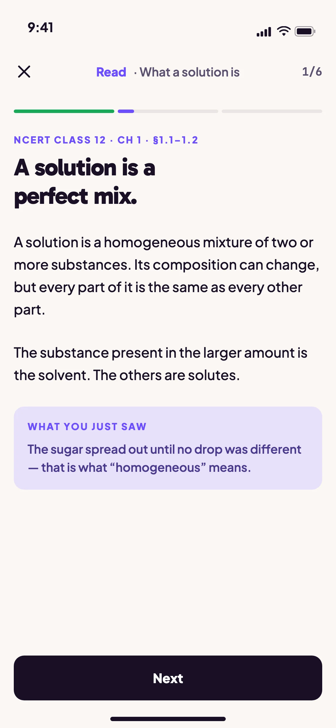
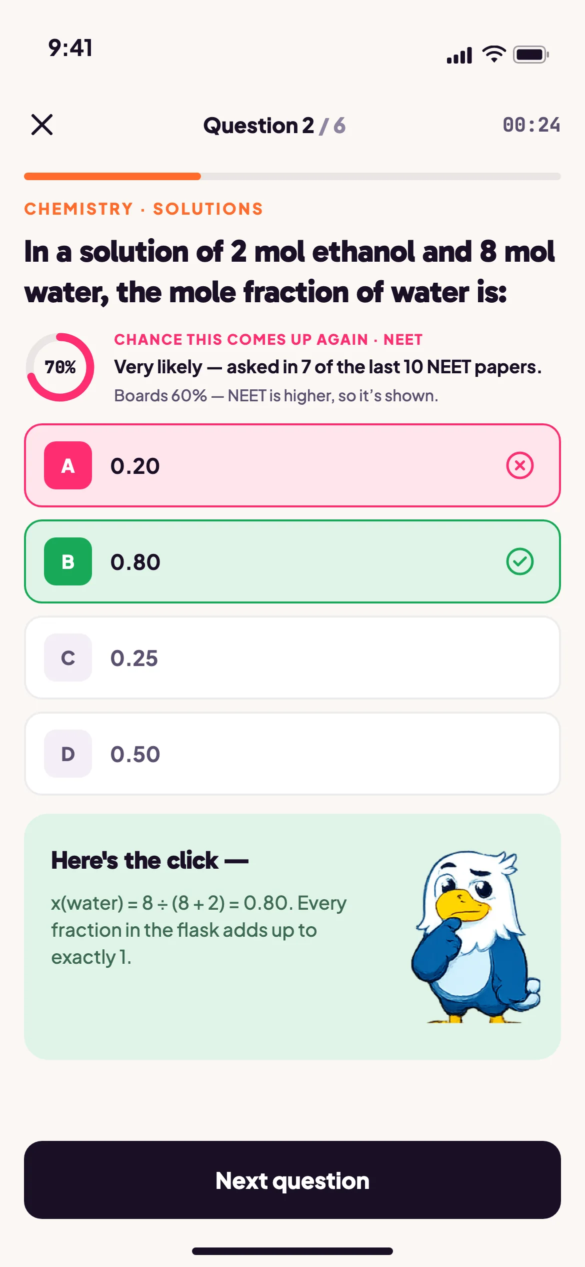
Reading a chapter and understanding it are different things. In the app every chapter is Do · Read · Verify: a game you play, a short read, then questions that check you. A full chapter, understood, in 45 to 60 minutes.
- DoA game built for the idea
- ReadThe short NCERT explainer
- VerifyQuestions that check you

Still stuck at 11pm? TarQ AI reads the answer you got wrong and teaches the idea behind it, not just the right option.